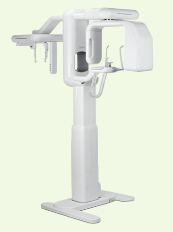
Full mouth X Ray & TMJ X Ray
A full mouth X-ray, also known as a panoramic X-ray or orthopantomogram (OPG), is a dental radiograph that provides a comprehensive view of a patient's teeth, jaws, and surrounding structures. It is an essential tool to diagnose and treat dental problems, such as cavities, gum disease, impacted teeth, and jaw disorders.
The process involves the patient standing or sitting still while a machine rotates around their head, capturing images of the upper and lower jaws, teeth, and surrounding bone structures. The image produced is a flat, two-dimensional view that displays the entire mouth in one shot, allowing the dentist to assess all of the teeth and jaw structures in a single image.
Full mouth X-rays are particularly useful for patients with complex dental problems or for those who are undergoing extensive dental work, such as full-mouth restorations or orthodontic treatment. They are also used to assess the growth and development of teeth in children and to detect any abnormalities in the jawbone or surrounding structures.

A full mouth X-ray provides a comprehensive view of the teeth, jawbones, and surrounding structures. It is often necessary for diagnosing and treating dental problems, such as cavities, gum disease, and impacted teeth. It is also helpful for planning treatments like orthodontic treatment or full-mouth restorations.
Yes, a full mouth X-ray is generally safe. It uses a low dose of radiation, and dental professionals take steps to minimize exposure. However, if you are pregnant or suspect you may be, inform your dentist before the X-ray, as radiation exposure can be harmful to a developing fetus.
The procedure typically takes less than 10 minutes, and the results are usually available immediately.
There is no special preparation required for a full mouth X-ray. You may be asked to remove any metal objects or jewelry from your head and neck area.
The frequency of full mouth X-rays varies depending on the patient's dental health and risk of developing dental problems. The dentist will determine how often you need this type of X-ray based on your individual needs.
A TMJ (temporomandibular joint) X-ray, also known as a TMJ radiograph, is a specialized dental X-ray that provides a detailed view of the temporomandibular joint. This joint connects the jawbone to the skull and is responsible for the movements of the jaw when speaking, chewing, and opening and closing the mouth.
TMJ X-rays are used to diagnose and evaluate the severity of TMJ disorders, which can cause pain, clicking, popping, and limited movement in the jaw. The X-ray images show the position and condition of the joint, including the shape and size of the joint's articular surfaces, the position of the condyle, and any signs of degeneration or damage to the joint.
The procedure involves the patient opening their mouth wide and positioning their head in a specific way to capture images of the joint. The images are typically taken in both the open and closed positions to assess the joint's movement and function.
A TMJ X-ray is a specialized dental X-ray that provides a detailed view of the temporomandibular joint. It is used to diagnose and evaluate the severity of TMJ disorders.
If you are experiencing pain, clicking, or popping in your jaw, a TMJ X-ray can help diagnose the cause of these symptoms. It can also show any damage or degeneration to the temporomandibular joint.
Yes, a TMJ X-ray is generally safe. It uses a low dose of radiation, and dental professionals take steps to minimize exposure. However, if you are pregnant or suspect you may be, inform the dentist before the X-ray, as radiation exposure can be harmful to a developing fetus.
The procedure typically takes less than 10 minutes, and the results are usually available immediately.
There is no special preparation required for a TMJ X-ray. You may be asked to remove any metal objects or jewelry from your head and neck area. The dentist may also ask you to open and close your mouth in various positions during the X-ray.
In summary, both full mouth X-rays and TMJ X-rays are important diagnostic tools used in dentistry to evaluate and treat dental problems. While full mouth X-rays provide a comprehensive view of the teeth and jaw structures, TMJ X-rays focus specifically on the temporomandibular joint and are used to diagnose TMJ disorders. Both X-ray types are safe and effective methods for evaluating dental health and should be used when clinically indicated.
If there's anything you'd like to know more about, just let us know - we're always happy to provide additional information
Fill the details will get back to you shortly...


Working Hours
Monday - Saturday : 10:30 AM - 7:00 PM
Thursday: Holiday
Sunday: 10:30 AM - 1:00 PM
Call Us
+91 91081 24151
+91 99001 14151
Mail Us
happytohelp@vatsandparam.com